|
|
|||||
|
||||||
|
||||||
|
|
|||||
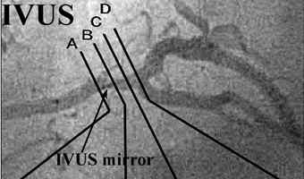
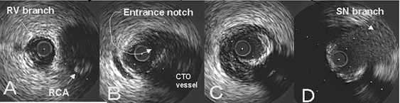
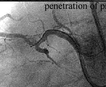
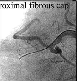
Figure 20: Locating the CTO ostium with IVUS
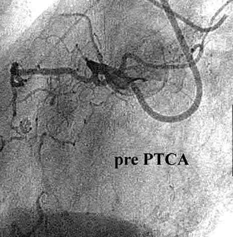
In this patient, the proximal RCA has a very tight bend and with CAG it is very
hard to get a good view of the area where the right ventricular branch forks
from the main trunk. As you pull back the IVUS catheter and the mirror, the
images obtained allow you to locate the CTO ostium.