|
|
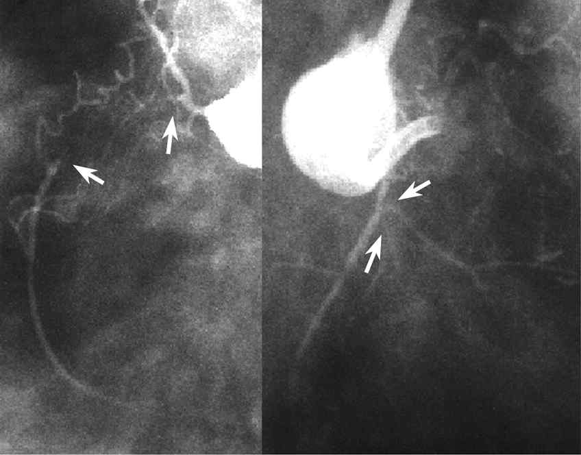
Figure 18: Case Demonstrating Side-branch Technique
You can see from this RAO view (b) that a relatively large right ventricular
branch forks at quite a wide angle. This large side-branch is not suitable for
the side-branch technique but the smaller right ventricular branch leading from
the same portion is suitable (c). A large dissection can be seen in the occluded
portion, but dilating with a 1.5mm balloon and consequent recanalization allows
the wire to pass along to the distal true lumen.