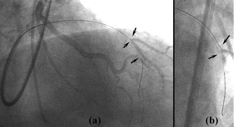
Figure 15: Breaking through the distal fibrous cap using the parallel wire
technique
The first wire passes along a false lumen and into a septal branch branching
off from the distal true lumen, and an attempt is made to cross the distal fibrous
cap with the second wire. At this point, from (a) the RAO + cranial view, it
looks wrongly as if the wire-tip is already in the true lumen. From (b), the
AP + cranial view, you can see that the true lumen is being stretched and widened
by the first wire.